Pressure ulcers develop when there is a prolonged reduction in blood flow to the layers of the skin which subsequently leads to skin tissue break down. Pressures above capillary pressure, >32-35mmHg for a period longer than 2 hours have been demonstrated to lead to capillary occlusion and ischemia (Daniel, Priest & Wheatley, 1981).
However, more recently it has been recognised that vulnerable patients may be susceptible to skin tissue damage after as little as 20 minutes of pressure exposure (Angmorterh, S.K., 2016; Moore & Patton, 2019). Whilst interventions to reduce the risk of pressure ulcers including high specification mattresses, pressure redistribution aids and offloading equipment have been considered in relation to pressure relief on hospital wards, the medical imaging context has only more recently been considered in relation to pressure ulcer risk (Alresheedi, Walton, Tootell, Webb & Hogg, 2020; Angmorterh, 2016).
Medical Imaging covers a broad range of procedures which vary in time frame. A patient may be in position for as little as 10 minutes (for example a peripheral limb X-ray), or 40-60 minutes for Radiotherapy planning, or at the upper end for 2 hours or more for interventional procedures, such as stent insertion.
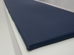
Whilst bed mattresses tend to be relatively thick and offer pressure relieving properties, X-ray mattresses are typically 2.5-5cm thick and therefore relatively high interface pressures have been recorded for patient equivalent weights on X-ray mattresses (Alresheedi et al., 2020).
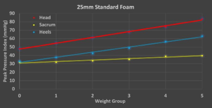
Peak Pressure Index - 25mm standard foam
Peak Pressure Index for the jeopardy areas, head (red), sacrum (yellow) and heels (blue). Weight groups are representative of a range of human equivalent body masses ranging from approximately 50 kg (Weight Group 0) up to 150 kg (Weight Group 5).


![]() and then Add to Home Screen.
and then Add to Home Screen.![[FP47] 25 mm X-Ray Table Mattress - Standard Foam - Sewn Cover](/web/image/product.product/74933/image_1024/%5BFP47%5D%20%5BFP47%5D%2025%20mm%20X-Ray%20Table%20Mattress%20-%20Standard%20Foam%20-%20Sewn%20Cover%20%28Navy%29?unique=29dcafb)


